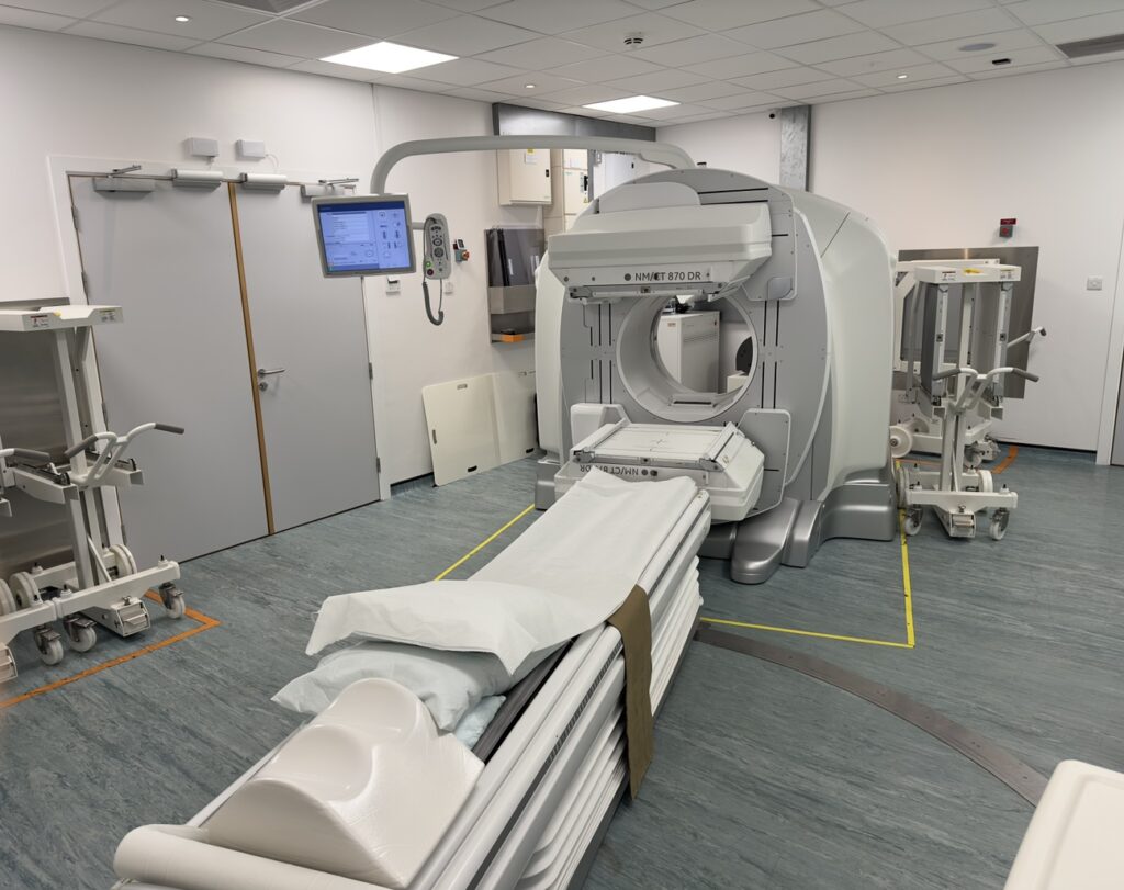
Most of the diagnostic nuclear medicine procedures involve imaging with a gamma camera and the Trust has 3 gamma cameras, two at Castle Hill Hospital and one at Hull Royal Infirmary.
Dual Headed Gamma Camera
A lot of our scans are performed on a dual-headed gamma camera. This means there are two detectors to acquire scan results.
Our dual-headed gamma cameras perform a range of different scans, depending on the test being performed:
- Static – the camera will be positioned over one or more areas of your body
- Dynamic – the camera acquires a series of images to show function over time, enabling visualisation of anatomy in motion
- Whole body – the camera slowly scans you from head to toe
- SPECT – the camera slowly rotates around area(s) of your body
- SPECT/CT – following a SPECT scan, a quick CT scan is acquired.

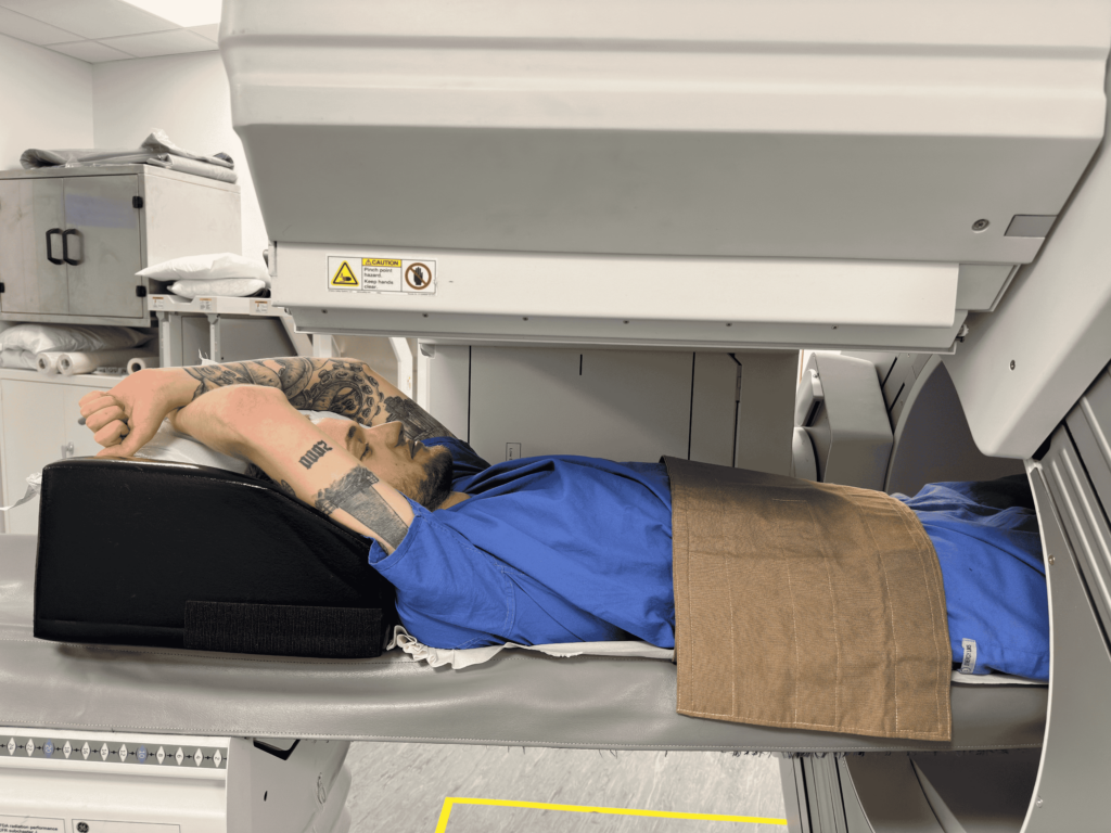
An immobilisation strap and knee cushion are used to ensure comfort and reduce movement during the scan. . If you would like the radio on or off during your scan, then please let a technologist know. Our technologists will always be observing from the control room.
Our detectors will come close to you, to obtain the best images possible. For whole body scans, your head is the first thing to be scanned, and should be clear of the detectors within the first 5-10 minutes. Below shows whole body positioning under the detectors.
Some scans (mainly heart and some SPECT/CT scans) may require arms to be raised for the duration of the scan, as shown below. This is to make it easier for the camera to get good-quality pictures.
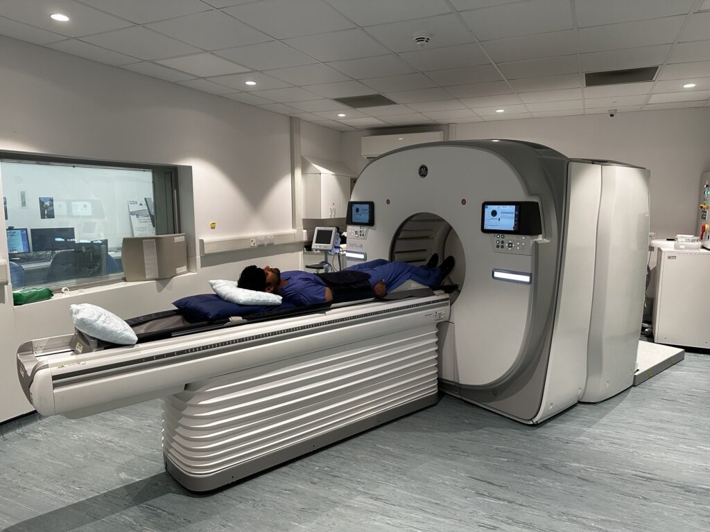
Multi-Detector CZT Camera (StarGuide)
This camera has 12 detectors that allow for faster SPECT/CT imaging than compared to our dual-headed gamma cameras but it cannot be used for all scan types.

An immobilisation strap and knee cushion are used to ensure comfort and reduce movement during the scan.. The detectors do move during the acquisition to capture all possible angles of your body. Our technologists will always be observing from the control room.
Generally, there is no need to wait in the department after your scan but a technologist will let you know otherwise. The images are reported by our radiologists or cardiologists, with the results being sent back to your consultant/care provider. Unfortunately, our technologists cannot discuss scan results or show you the images.
Images
This is a whole-body bone scan, the most commonly performed Nuclear Medicine procedure.

This is a three-dimensional image of the left ventricle of a heart. By looking at the heart in this way we can more easily see areas which are not working properly.

State-of-the-art imaging allows Nuclear Medicine and CT images to be taken during the same scan. By superimposing these images, we can more accurately see the location of any areas of disease.

